

We use Heidelberg Spectralis OCT which provides high-resolution spectral-domain optical coherence tomography, enabling detailed cross-sectional visualization of the retinal microstructure, macula and optic nerve head. With additional capabilities including OCT Angiography (OCTA), Fundus Fluorescein Angiography (FFA) and Indocyanine Green Angiography (ICG), the system facilitates comprehensive assessment of retinal and choroidal vascular pathology.
Services
Eye Treatments
Our specialists can advise on suitable clinical services during your consultation by combining state-of-the-art equipment with innovative treatment methods and therapeutic approaches. Many eye procedures are performed onsite in our modern, purpose-built treatment rooms at Strathfield Retina Clinic, while others are performed by our doctors at external facilities.
Eyelid & Eye Surface Treatments
Vitreo-Retinal Surgery
Equipment & Technology
Strathfield Retina Clinic continually invests in advanced medical equipment and technology to support the accurate diagnosis and treatment of eye conditions. Our high-resolution imaging systems and latest-generation laser technology enable detailed assessment, early detection and precise treatment of retinal disease, supporting optimal patient care and visual outcomes.

Retinal Scans and Imaging
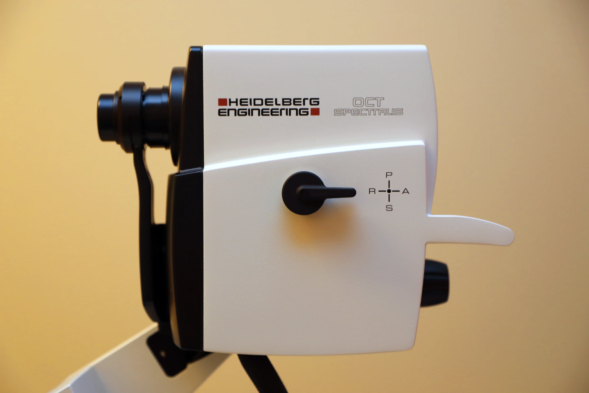
Advanced follow-up registration capabilities further facilitate assessment of retinal microvascular changes and precise longitudinal comparison of structural progression.
Optos ultra-widefield imaging system provides extensive visualization of the posterior pole and peripheral retina, capturing significantly more retinal surface area than conventional fundus photography.
This is particularly valuable in assessing peripheral retinal pathology, including retinal breaks, lattice degeneration, peripheral vascular abnormalities, haemorrhages and inflammatory changes. Our system incorporates advanced multimodal imaging, including fundus autofluorescence (FAF), FFA and ICG, enabling detailed evaluation of retinal and choroidal structural and vascular abnormalities.
Angiography

Our angiographic imaging capabilities include both FFA & ICG, providing complementary assessment of retinal and choroidal vasculature.
FFA enables dynamic evaluation of retinal vascular perfusion, capillary non-perfusion, vascular leakage, staining and neovascularization.
ICG angiography offers enhanced visualization of the choroidal circulation, particularly useful in the investigation of choroidal neovascularization, polypoidal choroidal vasculopathy and other choroidal vascular disorders.
Advanced Optical Biometry
For patients undergoing preoperative cataract assessment, we utilize the ZEISS IOL Master 700, which incorporates swept-source OCT technology to deliver high-precision, non-contact ocular biometry and detailed visualization of ocular structures. It provides accurate measurement of key biometric parameters for reliable intraocular lens (IOL) power calculations.
Its advanced imaging and fixation verification capabilities further enhance measurement reliability and assist in identifying anatomical variations that may influence surgical planning.
B-Scan Ultrasound
When direct visualization of the fundus is limited, such as in the presence of dense cataracts, vitreous haemorrhage or other media opacities, ophthalmic B-scan helps to provide cross-sectional imaging of the posterior segment.
This non-invasive imaging modality enables assessment of the vitreous, retina, choroid and optic nerve, assisting in the detection of retinal detachment, vitreous abnormalities, intraocular masses and other posterior segment pathologies when direct fundus examination is limited.
Visual Field Test
Field Analyzer is an essential diagnostic tool for detecting and monitoring glaucomatous visual field defects, optic neuropathies and other neurological or retinal conditions. With various testing strategies, ZEISS Humphrey Field Analyzer enables detailed assessment of visual field sensitivity, defect patterns and progression.
It also facilitates visual field assessment for patients requiring functional vision evaluation as part of driving license medical assessments and renewal requirements.
MAIA Microperimeter
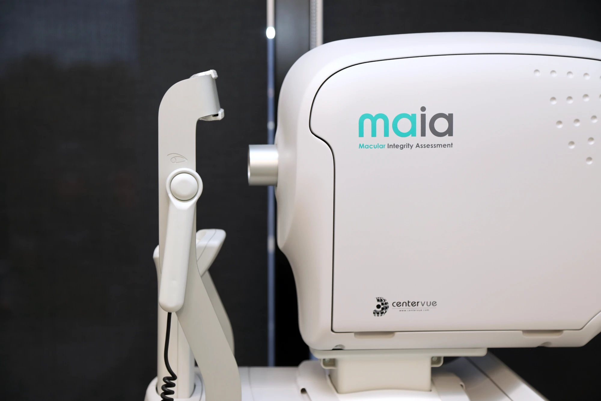
The MAIA (Macular Integrity Assessment) Microperimeter provides precise assessment of macular function through fundus-tracked microperimetry, correlating retinal sensitivity with corresponding fundus anatomy.
It enables detailed evaluation of central visual function, fixation stability and macular sensitivity, making it particularly useful in the assessment and monitoring of macular diseases, including age-related macular degeneration (AMD), macular dystrophies and other retinal disorders, complementing structural assessment with OCT imaging.