Digital Eye Scan
Good quality retinal photographs are integral in the diagnosis, treatment and progress monitoring of retinal disorders like diabetic retinopathy and macular degeneration. The clinical usefulness of a image is highly dependent on its quality. Using our high resolution digital cameras, excellent images are taken for every patient by our retinal trained orthoptists.
Anterior/Posterior Segment Digital Camera
Our Haag-Streit high resolution anterior/posterior segment digital eye scan can help diagnose and track disorders in the front, middle and back parts of the eye, including inflammatory conditions of the eye and far peripheral disorders of the retina and choroid.
Optical Coherence Tomography
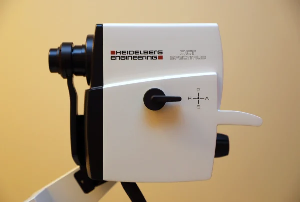
The Optical Coherence Tomography (OCT) digital eye scan at Strathfield Retina Clinic is the highest resolution laser scanner available with a precision eye-tracking device. This allows for earlier detection of sight-threatening retinal conditions.
Optical Coherence Tomography is a non-invasive digital eye scan of the retina which uses light to produce a detailed image of the retina and macula and is an essential instrument to diagnose and treat conditions such as Macular Degeneration, Diabetic Retinopathy, Retinal Vein Occlusion, Central Serous Retinopathy, Epiretinal Membrane and Vitreo-Macular Traction Syndrome. This highly sophisticated imaging tool takes slices of the retina and optic nerve and can produce a cross-sectional view of these structures and also detailed 3D reconstructions.
There is no radiation involved and the test is safe during pregnancy.
We have 2 different High Resolution Spectral Domain Optical Coherence Tomography machines, including the latest Eye-Tracking Scanning Laser Ophthalmoscope SPECTRALIS HRA + OCT and the SPECTRALIS OCT both from Heidelberg Engineering.
OCT 2 Angiography
Heidelberg Engineering have developed the Next Generation of OCT2 beyond standard Spectral Domain OCT which provides higher resolution scans, enhanced visualization of retinal, vitreal and choroidal structures and faster acquisition times.
OCT2 Angiography is the latest rapid non-invasive method of visualization of the retinal and choroidal vasculature without the need for dye injection. In combination with other imaging modalities, these advances in imaging often enable more accurate, less invasive diagnoses of eye disorders and a more comprehensive understanding of vascular abnormalities.